Friday 27 May 2011

VECTOR


INTRODUCTION TO VECTOR


DEFINITION
                        ‘’A vector is a DNA molecule used as a vehicle to transfer foreign genetic material into another cell.’’
The vector itself is generally a DNA sequence that consists of an insert (transgene) and a larger sequence that serves as the "backbone" of the vector.
A cloning vector is a small piece of DNA into which a foreign DNA fragment can be inserted. The insertion of the fragment into the cloning vector is carried out by treating the vehicle and the foreign DNA with a restriction enzyme that creates the same overhang, then ligating the fragments together. There are many types of cloning vectors. Genetically engineered plasmids and bacteriophages (such as phage λ) are perhaps most commonly used for this purpose. Other types of cloning vectors include bacterial artificial chromosomes (BACs) and yeast artificial chromosomes (YACs).
Purpose of a vector
 The purpose of a vector which transfers genetic information to another cell is typically to isolate, multiply, or express the insert in the target cell. 
To allow for convenient and favorable insertions, most cloning vectors have had nearly all their restriction sites engineered out of them and a synthetic multiple cloning site (MCS) inserted that contains many restriction sites. MCSs allow for insertions of DNA into the vector to be targeted and possibly directed in a chosen orientation. A selectable marker, such as an antibiotic resistance [e.g. beta-lactamase (see figure)] is often carried by the vector to allow the selection of positively transformed cells (see Screening below). All plasmids must carry a functional origin of replication (ORI; not shown in figure).
The four major types of vectors are plasmids, viruses, cosmids, and artificial chromosomes.
Common to all engineered vectors are an origin of replication, a multicloning site, and a selectable marker

Origin of replication

 The origin of replication (also called the replication origin) is a particular sequence in a genome at which replication is initiated. This can either be DNA replication in living organisms such as prokaryotes and eukaryotes, or RNA replication in RNA viruses, such as double-stranded RNA viruses. DNA replication may proceed from this point bidirectionally or unidirectionally.
The specific structure of the origin of replication varies somewhat from species to species, but all share some common characteristics such as high AT content. The origin of replication binds the pre-replication complex, a protein complex that recognizes, unwinds, and begins to copy DNA.

Types

The two types of replication origin are :
  • Narrow or broad host range
  • High- or low-copy number
There are also significant differences between prokaryotic and eukaryotic origins of replication:
  • Bacteria have a single circular molecule of DNA, and typically only a single origin of replication per circular chromosome.
  • Archaea have a single circular molecule of DNA, and several origins of replication along this circular chromosome.
  • Eukaryotes often have multiple origins of replication on each linear chromosome that initiate at different times (replication timing), with up to 100,000 present in a single human cell.Having many origins of replication helps to speed the duplication of their (usually) much larger store of genetic material. The segment of DNA that is copied starting from each unique replication origin is called a replicon.
Multiple cloning site
A multiple cloning site (MCS), also called a polylinker, is a short segment of DNA which contains many (up to ~20) restriction sites - a standard feature of engineered plasmids.Restriction sites within an MCS are typically unique, occurring only once within a given plasmid. MCSs are commonly used during procedures involving molecular cloning or subcloning. Extremely useful in biotechnology, bioengineering, and molecular genetics, MCSs let a biotechnologist insert a piece of DNA or several pieces of DNA into the region of the MCS. This can be used to create transgenic organisms, also known as genetically modified organisms (GMOs).
EXAMPLE
 The pUC18 and pUC19 polylinker

Selectable marker

 A selectable marker is a gene introduced into a cell, especially a bacterium or to cells in culture, that confers a trait suitable for artificial selection.
They are a type of reporter gene used in laboratory microbiology, molecular biology, and genetic engineering to indicate the success of a transfection or other procedure meant to introduce foreign DNA into a cell.
Selectable markers are often antibiotic resistance genes; bacteria that have been subjected to a procedure to introduce foreign DNA are grown on a medium containing an antibiotic, and those bacterial colonies that can grow have successfully taken up and expressed the introduced genetic material.
Examples of selectable markers include:the Abicr gene
Common vectors
Two common vectors are plasmids and viral vectors.

Plamid

A plasmid is a DNA molecule that is separate from, and can replicate independently of, the chromosomal DNA.. They are double-stranded and, in many cases, circular.
Plasmids usually occur naturally in bacteria, but are sometimes found in eukaryotic organisms (e.g., the 2-micrometre-ring in Saccharomyces cerevisiae).Plasmid sizes vary from 1 to over 1,000 kilobase pairs (kbp). The number of identical plasmids in a single cell can range anywhere from one to even thousands under some circumstances.
The term plasmid was first introduced by the American molecular biologist Joshua Lederberg in 1952.
Illustration of a bacterium with plasmid enclosed showing chromosomal DNA and plasmid
Plasmids are considered transferable genetic elements, or "replicons", capable of autonomous replication within a suitable host.
Plasmids can be found in all three major domains: Archea, Bacteria and Eukarya.
Unlike viruses, plasmids are "naked" DNA and do not encode genes necessary to encase the genetic material for transfer to a new host, though some classes of plasmids encode the sex pilus necessary for their own transfer. Plasmid host-to-host transfer requires direct, mechanical transfer by conjugation or changes in host gene expression allowing the intentional uptake of the genetic element by transformation.
 Plasmids may carry genes that provide resistance to naturally occurring antibiotics in a competitive environmental niche, or alternatively the proteins produced may act as toxins under similar circumstances. Plasmids also can provide bacteria with an ability to fix elemental nitrogen or to degrade recalcitrant organic compounds which provide an advantage when nutrients are scarce.
There are two types of plasmid integration into a host bacteria: Non-integrating plasmids replicate as with the top instance; whereas episomes, the lower example, integrate into the host chromosome.
However, a plasmid can only contain inserts of about 1–10 kbp. To clone longer lengths of DNA, lambda phage with lysogeny genes deleted, cosmids, bacterial artificial chromosomes or yeast artificial chromosomes could be used.
CONSTRUCTION OF  PLASMID VECTOR.
Plasmid can be good cloning vector because they carry an origin of replication and are therefore able to replicate independently within cell.
Most plasmid used as vectors also encode some type of selectable marker such as gene of resistance as amphiciline.
If  the host cell are amphiciline sensitive,the only host cell can grow on medium containing amphiciline are thosethat have taken up plasmid.
Vector must also havue a small sequence of base pair that can be recognized by restriction enzyme.when this enzyme open the circular plasmid ,foreign DNA can be incorporated.
When plasmid vector and foreign DNA are both cut with same restriction enzyme and mixed together, not all molecule will join to form recombinant.
Some vector molecule will reaneal without incorporating foreign DNA .To identify cells that contain plasmids have incorporated foreign DNA a second marker gene is needed on the vector.

Episomes

An episome is a portion of genetic material that can exist independent of the main body of genetic material (called the chromosome) at some times, while at other times is able to integrate into the chromosome.Examples of episomes include insertion sequences and transposons.
Another way to classify plasmids is by function. There are five main classes:
  • Fertility-F-plasmids, which contain tra-genes. They are capable of conjugation (transfer of genetic material between bacteria which are touching).
  • Resistance-(R)plasmids, which contain genes that can build a resistance against antibiotics or poisons and help bacteria produce pili. Historically known as R-factors, before the nature of plasmids was understood.
  • Col-plasmids, which contain genes that code for (determine the production of) bacteriocins, proteins that can kill other bacteria.
  • Degradative plasmids, which enable the digestion of unusual substances, e.g., toluene or salicylic acid.
  • Virulence plasmids, which turn the bacterium into a pathogen (one that causes disease).

Yeast plasmids

Other types of plasmids are often related to yeast cloning vectors that include:
  • Yeast integrative plasmid (YIp), yeast vectors that rely on integration into the host chromosome for survival and replication, and are usually used when studying the functionality of a solo gene or when the gene is toxic. Also connected with the gene URA3, that codes an enzyme related to the biosynthesis of pyrimidine nucleotides (T, C);
  • Yeast Replicative Plasmid (YRp), which transport a sequence of chromosomal DNA that includes an origin of replication. These plasmids are less stable, as they can "get lost" during the budding.
 Viral vector
Viral vectors are a tool commonly used by molecular biologists to deliver genetic material into cells. This process can be performed inside a living organism (in vivo) or in cell culture (in vitro). Viruses have evolved specialized molecular mechanisms to efficiently transport their genomes inside the cells they infect. Delivery of genes by a virus is termed transduction and the infected cells are described as transduced. Molecular biologists first harnessed this machinery in the 1970s. Paul Berg used a modified SV40 virus containing DNA from the bacteriophage lambda to infect monkey kidney cells maintained in culture.

Key properties of a viral vector

Viral vectors are tailored to their specific applications but generally share a few key properties.
  • Safety: Although viral vectors are occasionally created from pathogenic viruses, they are modified in such a way as to minimize the risk of handling them. This usually involves the deletion of a part of the viral genome critical for viral replication. Such a virus can efficiently infect cells but, once the infection has taken place, requires a helper virus to provide the missing proteins for production of new virions.
  • Low toxicity: The viral vector should have a minimal effect on the physiology of the cell it infects.
  • Stability: Some viruses are genetically unstable and can rapidly rearrange their genomes. This is detrimental to predictability and reproducibility of the work conducted using a viral vector and is avoided in their design.
  • Cell type specificity: Most viral vectors are engineered to infect as wide a range of cell types as possible. However, sometimes the opposite is preferred. The viral
 Types of viral vectors

Retroviruses

 Retroviruses are one of the mainstays of current gene therapy approaches. The recombinant retroviruses such as the Moloney murine leukemia virus have the ability to integrate into the host genome in a stable fashion. They contain a reverse transcriptase that allows integration into the host genome..
The primary drawback to use of retroviruses such as the Moloney retrovirus involves the requirement for cells to be actively dividing for transduction. As a result, cells such as neurons are very resistant to infection and transduction by retroviruses. There is concern that insertional mutagenesis due to integration into the host genome might lead to cancer or leukemia.
Lentiviruses
Lentiviruses are a subclass of Retroviruses. They have recently been adapted as gene delivery vehicles (vectors) thanks to their ability to integrate into the genome of non-dividing cells, which is the unique feature of Lentiviruses as other Retroviruses can infect only dividing cells. The viral genome in the form of RNA is reverse-transcribed when the virus enters the cell to produce DNA, which is then inserted into the genome at a random position by the viral integrase enzyme. The vector, now called a provirus, remains in the genome and is passed on to the progeny of the cell when it divides.

Adenoviruses

Adenoviral DNA does not integrate into the genome and is not replicated during cell division. This limits their use in basic research, although adenoviral vectors are occasionally used in in vitro experiments. Their primary applications are in gene therapy and vaccination. Since humans commonly come in contact with adenoviruses, which cause respiratory, gastrointestinal and eye infections, they trigger a rapid immune response with potentially dangerous consequences. To overcome this problem scientists are currently investigating adenoviruses to which humans do not have immunity.

Adeno-associated viruses

Adeno-associated virus (AAV) is a small virus that infects humans and some other primate species. AAV is not currently known to cause disease and consequently the virus causes a very mild immune response. AAV can infect both dividing and non-dividing cells and may incorporate its genome into that of the host cell. These features make AAV a very attractive candidate for creating viral vectors for gene therapy.[1]

Nanoengineered substances

Nonviral substances such as Ormosil have been used as DNA vectors and can deliver DNA loads to specifically targeted cells in living animals. (Ormosil stands for organically modified silica or silicate.)

Expression vector and its construction

An expression vector, otherwise known as an expression construct, is generally a plasmid that is used to introduce a specific gene into a target cell. Once the expression vector is inside the cell, the protein that is encoded by the gene is produced by the cellular-transcription and translation machinery ribosomal complexes. The plasmid is frequently engineered to contain regulatory sequences that act as enhancer and promoter regions and lead to efficient transcription of the gene carried on the expression vector.The goal of a well-designed expression vector is the production of large amounts of stable messenger RNA, and therefore proteins. Expression vectors are basic tools for biotechnology and the production of proteins such as insulin that are important for medical treatments of specific diseases like diabetes.
Expression vectors are used for molecular biology techniques such as site-directed mutagenesis. Cloning vectors, which are very similar to expression vectors, involve the same process of introducing a new gene into a plasmid, but the plasmid is then added into bacteria for replication purposes. In general, DNA vectors that are used in many molecular-biology gene-cloning experiments need not result in the expression of a protein.

EXPRESSION VECTOR.

Shuttle vector

 A shuttle vector is a vector (usually a plasmid) constructed so that it can propagate in two different host species [1]. Therefore, DNA inserted into a shuttle vector can be tested or manipulated in two different cell types. The main advantage of these vectors is they can be manipulated in E. coli then used in a system which is more difficult or slower to use (e.g. yeast, other bacteria).
Shuttle vectors include plasmids that can propagate in eukaryotes and prokaryotes (e.g. both Saccharomyces cerevisiae and Escherichia coli) or in different species of bacteria (e.g. both E. coli and Rhodococcus erythropolis). There are also adenovirus shuttle vectors, which can propagate in E. coli and mammals.Shuttle vectors are frequently used to quickly make multiple copies of the gene in E. coli (amplification). They can also be used for in vitro experiments and modifications (e.g. mutagenesis, PCR)

Monday 16 May 2011

Ozone and the ozone layer

The Ozone Layer

The ozone layer is a layer in Earth's atmosphere which contains relatively high concentrations of ozone (O3). This layer absorbs 97-99% of the sun's high frequency ultraviolet light, which is potentially damaging to life on earth. Over 90% of ozone in earth's atmosphere is present here "Relatively high" means a few parts per million—much higher than the concentrations in the lower atmosphere but still small compared to the main components of the atmosphere. It is mainly located in the lower portion of the stratosphere from approximately 15 km to 35 km above Earth's surface, though the thickness varies seasonally and geographically.

A dobson unit is the most basic measure used in ozone research.One Dobson Unit (DU) is defined to be 0.01 mm thickness at STP (standard temperature and pressure).
Ozone layer thickness is expressed in terms of Dobson units, which measure what its physical thickness would be if compressed in the Earth's atmosphere. In those terms, it's very thin indeed. A normal range is 300 to 500 Dobson units, which translates to an eighth of an inch-basically two stacked pennies.

Dobson Unit


In space, it's best not to envision the ozone layer as a distinct, measurable band. Instead, think of it in terms of parts per million concentrations in the stratosphere (the layer six to 30 miles above the Earth's surface).

The unit is named after G.M.B. Dobson, one of the first scientists to investigate atmospheric ozone .

A thinning ozone layer leads to a number of serious health risks for humans. It causes greater incidences of skin cancer and cataract of the eye, with children being particularly vulnerable. There are also serious impacts for biodiversity. Increased UV-B rays reduce levels of plankton in the oceans and subsequently diminish fish stocks. It can also have adverse effects on plant growth, thus reducing agricultural productivity. Another negative effect is the reduced lifespan of certain materials.

Severe depletion of the Antarctic ozone layer was first observed in the early 1980s. The international response embodied in the Montreal Protocol. Today 191 countries worldwide have signed the Montreal Protocol which is widely regarded as the most successful Multinational Environmental Agreement ever reached to date.

Furthermore the phasing out of ozone depleting substances (ODS) has helped to fight climate change since many ODS are also powerful greenhouse gases.

HISTORY OF THE OZONE LAYER
600,000,000 B.C. Ozone layer forms
1839 Christian Schöenbein identifies ozone in the laboratory
1845 Auguste de la Rive and Jean-Charles de Marignac suggest ozone is a form of oxygen; confirmed by Thomas Andrews in 1856
1858 Andrei Houzeau finds ozone present in natural air
1865 Jean-Louis Soret proves that ozone is O3
1879 Marie Alfred Cornu measures solar spectrum and finds sharp cutoff in ultraviolet (UV) light
1881 Walter Hartley recognizes cutoff corresponds to UV absorption by ozone
1913 John William Strutt (Lord Rayleigh) shows absorption is not in lower atmosphere
1919 Charles Fabry makes first spectrometric measurements of "thickness" of ozone layer
1924 G.M.B. Dobson develops ozone spectrophotometer and begins regular measurements of ozone abundance (Arosa, Switzerland)
1925 Jean Cabannes and Jean Dufay show ozone is about 10 miles high
1928 Thomas Midgley synthesizes chlorofluorocarbons (CFC's)
1929 Umkehr method for Dobson instrument establishes that ozone maximum is below 15 miles altitude
1930 Sydney Chapman describes theory that explains existence of an ozone "layer"
1934 Ozonesonde (balloon) measurements establish the ozone concentration is maximum around 12 miles up
1930's GM develops applications for CFC's
1950 David Bates and Marcel Nicolet propose catalytic (HOx) ozone destruction
1957 Global network of Dobson spectrophotometers established during the International Geophysical Year (IGY)
late 1950's CFC market expands rapidly
early 1960's Catalytic destruction is necessary in order to explain ozone amounts
1960's Boeing proposes supersonic transport (SST) fleet of 800 aircraft
1969 Paul Crutzen discovers NOx catalytic cycle
1971-74 Dept of Transportation sponsors intensive program of research, The Climatic Impact Assessment Program (CIAP)
1971 Congress axes funding for the SST
1971 Johnston calculates that NOx from SST's could deplete ozone layer
1973 Rick Stolarski and Ralph Cicerone suggest catalytic capability of Cl
1973 James Lovelock detects CFC's in atmosphere
1974 Sherwood Rowland and Mario Molina warn of ozone depletion due to CFC's
March 1977 First international meeting (Washington DC) to address issue of ozone depletion held by the United Nations Environmental Programme (UNEP)
March 1978 US bans non-essential use of CFC's as aerosol propellant
1978 Total Ozone Mapping Spectrometer (TOMS) is launched aboard NIMBUS-7 spacecraft giving global coverage of ozone layer thickness
1980's Renewed expansion of CFC market
Oct 1982 Shigeru Chubachi measures low ozone over Syowa, Antarctica (reported at Ozone Commission meeting in Halkidiki, Greece in Sept 1984)
1984 British Antarctic Survey scientists discover recurring springtime Antarctic ozone hole (published in Nature May 1985)
March 1985 Vienna Convention for the Protection of the Ozone Layer
Sept. 1987 Montreal Protocol on Substances That Deplete the Ozone Layer (Amendments - London 1990; Copenhagen 1992)
March 1988 DuPont agrees to CFC production phase-out
late 1980's Ten years of satellite data begin to show measurable ozone depletion globally
1991 DuPont announces phase-out of CFC production by end of 1996
1992/3 Abnormally low ozone observed globally
1995 Crutzen, Rowland, and Molina win Nobel Prize in Chemistry
mid-1990's springtime Arctic ozone dent appearing
Jan. 1996 CFC production ends in US and Europe
2000 Maximum CFC concentrations in stratosphere are reached
Today The Ozone Layer - Global Map

THE FUTURE
2010 CFC production ends world-wide
2030 Hydrochlorofluorocarbon (HCFC) alternatives are phased out
2040 HCFC production ends world-wide
2050 Springtime Antarctic ozone hole disappears

Saturday 14 May 2011

History of Football

History of Football

A Brief History of the Game

Football’s Early Beginnings

Football (as well as rugby and soccer) are believed to have descended from the ancient Greek game of harpaston.   Harpaston is mentioned frequently in classical literature, where it is often referred to as a “very rough and brutal game“.  The rules of this ancient sport were quite simple:  Points were awarded when a player would cross a goal line by either kicking the ball, running with it across the goal line, or throwing it across the line to another player. The other team’s objective was simply to stop them by any means possible.  There was no specific field length, no side line boundaries, no specified number of players per team, only a glaring lack of rules.

Harpaston:  Luckily (for everyone) uniforms & equipment have improved dramatically.
Most modern versions of football are believed to have originated from England in the twelfth century. The game became so popular in England that the kings of that time (Henry II and Henry IV) actually banned football. They believed that football was taking away interest from the traditional sports of England, such as fencing and archery.

Evolution and the Beginnings of Standardization

Football didn’t really begin to take on any consistency of rules and boundaries until it was picked up as a sport in the seven major public schools of England in the early 1800’s.  Six of the seven schools were largely playing the same game (including Eton, Harrow and Winchester) - while the seventh, Rugby School (founded in 1567) was playing a markedly different version of football.
The other schools moved ahead refining their rules and eventually their game became known as "association football" – or soccer, which was played back then much as it is today.
Rugby School went in a different direction.  How and why the game developed differently at Rugby School appears to have been lost in history, but what is known is that by the 1830's, running with the ball at Rugby School was in common use and 18 foot goal posts had been added with a cross-bar at 10 feet above the ground.
The inclusion of the cross-bar was accompanied by a rule that a goal could only be scored by the ball passing over the bar from a place kick or drop kick. Apparently this was done to make scoring easier from further out and also to avoid the horde of defenders standing in and blocking the mouth of the goal.
Players who were able to "touch down" the ball behind the opponents goal line were awarded a "try-at-goal" - the player would make a mark on the goal line and then walk back onto the field of play to a point where a place kick at the goal was possible (a conversion). There was also an "off-your-side" rule used to keep the teams apart.  Passing the ball forward was not allowed.
By the mid-1860s British schools and universities had taken up Rugby's game and honored the school by giving the "new football" the name of rugby.
The game soon went trans-Atlantic to America and landed on fertile soil.

Roots of American Football

The birth date of football in the United States is generally regarded by football historians as November 6, 1869, when teams from Rutgers and Princeton Universities met for the first intercollegiate football game. In those early games, there were 20 players to a team and football still more closely resembled rugby than modern football.
The game of football has a history of constant rule changes. Rule changes have been implemented to bolster the excitement of the game of football and to increase the game's safety.
In 1873, representatives from Columbia, Rutgers, Princeton, and Yale Universities met in New York City to formulate the first intercollegiate football rules for the increasingly popular game. These four teams established the Intercollegiate Football Association (IFA) and set 15 as the number of players allowed on each team.
Walter Camp, the coach at Yale and a dissenter from the IFA over his desire for an eleven man team, helped begin the final step in the evolution from rugby-style play to the modern game of American football. The IFA’s rules committee, led by Camp, soon cut the number of players from fifteen to eleven, and also instituted the size of the playing field, at one hundred ten yards. In 1882 Camp also introduced the system of downs. After first allowing three attempts to advance the ball five yards, in 1906 the distance was changed to ten yards. The fourth down was added in 1912.
Within a decade, concern over the increasing brutality of the game led to its ban by some colleges. Nearly 180 players had suffered serious injuries, and eighteen deaths had been reported from the brutal mass plays that had become common practice. So in 1905, President Theodore Roosevelt called upon Harvard, Princeton, and Yale to help save the sport from demise.
At a meeting between the schools, reform was agreed upon, and at a second meeting, attended by more than sixty other schools, the group appointed a seven member Rules Committee and set up what would later become known as the National Collegiate Athletic Association, or the NCAA.
From this committee came the legalization of the forward pass, which resulted in a redesign of the ball and a more open style of play on the field. The rough mass plays, which once caused so many serious injuries, were prohibited by the committee. Also prohibited was the locking of arms by teammates in an effort to clear the way for their ball carriers. The length of the game was shortened, from seventy to sixty minutes, and the neutral zone, which separates the teams by the length of the ball before each play begins, was also established.

Fresh Fruit & Vegetables

Fresh Fruit & Vegetables

Enjoy a healthier lifestyle by eating
more fresh fruit & vegetables...     

Fresh Fruit & Vegetables  
Everyone agrees on the importance of eating more fresh fruits & vegetables, but not enough people are following this important advice. Dietary experts recommend that every person should eat at least five servings of fresh fruits & vegetables every day. Increasing your consumption of fruits and vegetables is one of the easiest changes you can make to increase your level of health, lose weight and gain fitness.

The latest food guidelines recommend that adults eat from five to nine servings of fresh fruit & vegetables every day. While that may seem like a lot, it is an important goal to strive for, and a very reachable one.

A serving of a fruit or vegetable is equal to:

1. 1 medium sized vegetable or fruit (such as an apple, orange or banana)

2. 2 small fruits (such as kiwi fruit or plums)

3. ½ cup of fresh, frozen or canned fruits or vegetables

4. ½ cup of 100% fruit juice

5. ¼ cup of dried fruit

6. 1 cup of green salad

Study after study has shown that a diet rich in fresh fruit & vegetables lowers the risk of certain cancers, heart disease and other chronic diseases and conditions. One reason for the recommendation that everyone increase their consumption of fruits and vegetables is that many of these foods have been shown to have strong antioxidant qualities.

Many fresh fruits & vegetables have high amounts of many antioxidant vitamins, including vitamin A, vitamin E and vitamin C. In addition to their importance as source of vitamins and minerals, fruits and vegetables also provide essential dietary fiber. 
 

Many people do not realize, that in addition to vitamin C, oranges, grapefruits and other citrus fruits also contain significant amounts of fiber. Fiber plays an important role in digestion, and it is thought to have protective qualities against heart disease and some forms of cancer. In addition, fiber is thought to have the ability to lower high levels of cholesterol in the blood.

Another great feature of fresh fruits & vegetables, especially to those watching their weight, is the high nutrition, low fat, and low calorie nature of these foods. Fruits and vegetables contain very low levels of fats, and a diet low in fat can be quite effective for long-term weight loss. In addition, fruits and vegetables contain no cholesterol, and they are lower in calories than many other types of foods.

Fresh fruits & vegetables have a lot of advantages besides just their nutritional importance. For one thing, they taste great and add a great deal of variety to everyday meals. Fruits and vegetables come in such a wide variety of colors, textures and flavors that they can be used in virtually every meal. Those seeking to maximize their consumption of fruits and vegetables should get into the habit of using fruits in salads, as toppings and as garnishes. 
 

No matter what your reason for following a healthy diet, we believe you will find that eating more fruits and vegetables is a delicious, as well as a nutritious, way to get the vitamins and minerals you need every day.

I hope that reading the above information was both enjoyable and educational for you. When word gets around about your command of healthy diet facts, others who need to know about fresh fruits & vegetables will start to actively seek you out.

What is a heart attack?


Picture of the heartWhat is a heart attack?

A heart attack (also known as a myocardial infarction) is the death of heart muscle from the sudden blockage of a coronary artery by a blood clot. Coronary arteries are blood vessels that supply the heart muscle with blood and oxygen. Blockage of a coronary artery deprives the heart muscle of blood and oxygen,causing injury to the heart muscle. Injury to the heart muscle causes chest pain and chest pressure sensation. If blood flow is not restored to the heart muscle within 20 to 40 minutes, irreversible death of the heart muscle will begin to occur. Muscle continues to die for six to eight hours at which time the heart attack usually is "complete." The dead heart muscle is eventually replaced by scar tissue.
Approximately one million Americans suffer a heart attack each year. Four hundred thousand of them die as a result of their heart attack.

What causes a heart attack? 


Atherosclerosis

Atherosclerosis is a gradual process by which plaques (collections) of cholesterol are deposited in the walls of arteries. Cholesterol plaques cause hardening of the arterial walls and narrowing of the inner channel (lumen) of the artery. Arteries that are narrowed by atherosclerosis cannot deliver enough blood to maintain normal function of the parts of the body they supply. For example, atherosclerosis of the arteries in the legs causes reduced blood flow to the legs. Reduced blood flow to the legs can lead to pain in the legs while walking or exercising, leg ulcers, or a delay in the healing of wounds to the legs. Atherosclerosis of the arteries that furnish blood to the brain can lead to vascular dementia (mental deterioration due to gradual death of brain tissue over many years) or stroke (sudden death of brain tissue).
In many people, atherosclerosis can remain silent (causing no symptoms or health problems) for years or decades. Atherosclerosis can begin as early as the teenage years, but symptoms or health problems usually do not arise until later in adulthood when the arterial narrowing becomes severe. Smoking cigarettes, high blood pressure, elevated cholesterol, and diabetes mellitus can accelerate atherosclerosis and lead to the earlier onset of symptoms and complications, particularly in those people who have a family history of early atherosclerosis.
Coronary atherosclerosis (or coronary artery disease) refers to the atherosclerosis that causes hardening and narrowing of the coronary arteries. Diseases caused by the reduced blood supply to the heart muscle from coronary atherosclerosis are called coronary heart diseases (CHD). Coronary heart diseases include heart attacks, sudden unexpected death, chest pain (angina), abnormal heart rhythms, and heart failure due to weakening of the heart muscle.
Atherosclerosis and angina pectoris

Angina pectoris (also referred to as angina) is chest pain or pressure that occurs when the blood and oxygen supply to the heart muscle cannot keep up with the needs of the muscle. When coronary arteries are narrowed by more than 50 to 70 percent, the arteries may not be able to increase the supply of blood to the heart muscle during exercise or other periods of high demand for oxygen. An insufficient supply of oxygen to the heart muscle causes angina. Angina that occurs with exercise or exertion is called exertional angina. In some patients, especially diabetics, the progressive decrease in blood flow to the heart may occur without any pain or with just shortness of breath or unusually early fatigue.
Exertional angina usually feels like a pressure, heaviness, squeezing, or aching across the chest. This pain may travel to the neck, jaw, arms, back, or even the teeth, and may be accompanied by shortness of breath, nausea, or a cold sweat. Exertional angina typically lasts from one to 15 minutes and is relieved by rest or by taking nitroglycerin by placing a tablet under the tongue. Both resting and nitroglycerin decrease the heart muscle's demand for oxygen, thus relieving angina. Exertional angina may be the first warning sign of advanced coronary artery disease. Chest pains that just last a few seconds rarely are due to coronary artery disease.
Angina also can occur at rest. Angina at rest more commonly indicates that a coronary artery has narrowed to such a critical degree that the heart is not receiving enough oxygen even at rest. Angina at rest infrequently may be due to spasm of a coronary artery (a condition called Prinzmetal's or variant angina). Unlike a heart attack, there is no permanent muscle damage with either exertional or rest angina.
Atherosclerosis and heart attack

Occasionally the surface of a cholesterol plaque in a coronary artery may rupture, and a blood clot forms on the surface of the plaque. The clot blocks the flow of blood through the artery and results in a heart attack (see picture below). The cause of rupture that leads to the formation of a clot is largely unknown, but contributing factors may include cigarette smoking or other nicotine exposure, elevated LDL cholesterol, elevated levels of blood catecholamines (adrenaline), high blood pressure, and other mechanical and biochemical forces.

Unlike exertional or rest angina, heart muscle dies during a heart attack and loss of the muscle is permanent, unless blood flow can be promptly restored, usually within one to six hours.
Heart Attack illustration - Myocardial Infarction
While heart attacks can occur at any time, more heart attacks occur between 4:00 A.M. and 10:00 A.M. because of the higher blood levels of adrenaline released from the adrenal glands during the morning hours. Increased adrenaline, as previously discussed, may contribute to rupture of cholesterol plaques.
Approximately 50% of patients who develop heart attacks have warning symptoms such as exertional angina or rest angina prior to their heart attacks, but these symptoms may be mild and discounted.

History and Uses of Sunglasses

History and Uses of Sunglasses

History


Sunglasses are used to aid your vision. Generally, it features darkened or colored lenses to protect the eyes from strong lights. It was said that Nero, a Roman Emperor watched fights of gladiators via polished gems, most likely emeralds.
The first usage of sunglasses was recorded during the 12th century in China. The lenses were made of smoky quarts flattened into panes. This does not provide corrective powers but the eyes are protected from glare. According to contemporary documents, the judges in Chinese Trial Courts used sunglasses to hide their facial expression while probing witnesses. This is similar on the Blind Justice representation in Western arts.
During the 18th century, James Ayscough started to experiment with colored lenses in eyeglasses. These are not considered as sunglasses because Ayscough believed that green or blue tinted glass corrects certain vision impairments. He was not concerned about the UV protection.
At the turn of the 1900s, wearing sunglasses became more popular and widespread in silent movies. The stars wear sunglasses not only to protect their eyes from the studio's harsh bright lighting but to avoid from being recognized.
In 1929, Sam Foster introduced mass-produced inexpensive sunglasses to America. He found ready markets on New Jersey and Atlantic City's beaches, selling his merchandise in the name of Foster Grant.
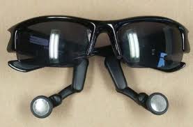
In 1936, sunglasses were polarized by Edwin H. Land using an original Polaroid filter. Oakley in 2004 created Thump. These sunglasses are equipped with a digital audio player that is built-in. Many smaller companies have copied this design.

Uses


• Hide the eyes during one on one communication. Typically, it hides weeping which is a sign of mourning, makes eye contacts impossible which is sometimes very intimidating, and shows detachment in some groups. Other reasons include hiding contracted or dilated pupils and bloodshot eyes (caused by drug use or physical abuse) and compensating for optimum photosensitivity.
• Fashion accessory. Wearing sunglasses always have a purpose. Darkened sunglasses in different shapes are a type of accessory. Compared to normal eyeglasses which can project a nerdy image, sunglasses are worn indoors or at night for projecting cool images and nonverbal communications.
• Hide serious visual impairments. It includes blindness, cross eyed condition, and abnormal appearance of the eyes because of cataract and nystagmus (uncontrollable jerking of the eyes). People wear sunglasses to prevent others from becoming uncomfortable.
• Visual comfort and clarity. Sunglasses protect the eyes from glare. Sometimes, a patient who received mydriatic eye drops is required to wear even disposable sunglasses after eye examinations.
• Protection. Prolonged exposure to UV rays causes long-term or short-term ocular problems including snow blindness, pterygium, photokeratitis, and some types of eye cancers. Medical experts revealed that wearing sunglasses is important for eye protection from UV rays. However, health authorities warn the public not to look at the sun during a solar eclipse using only sunglasses.
CE mark in the European Union identifies sunglasses that passed quality regulations. Keep in mind that there is also no connection between increased ultraviolet protection and high prices of sunglasses. Based on the 1995 study, polarizing sunglasses and expensive brands never guarantee a maximum UVA protection. The ACCC (Australian Competition and Consumer Commission) reports that consumers should never rely on prices as indicators of quality. Some unscientific survey revealed that there are generic sunglasses costing 6.95 dollars providing slightly better UV protection than Salvatore Ferragamo and Michael Kors shades. Oakley sunglasses have passed the requirements of the ANSI (American National Standards Institute) Z87.1 offering UV protection.
Most recently, HEV (high energy visible light) has been associated with macular degeneration related to age. Several manufacturers create sunglasses for blocking it. Children are also advised to wear UV protected sunglasses since their ocular lenses transmits significant amount of HEV light as compared to adults.
Some sunglasses even pass the requirements of ANSI Z87.1 for high impact and basic impact protection. However, compliance to these standards are voluntary. Therefore, not all manufacturers or sunglasses are required for compliance. Basically, the impact test is conducted by dropping a 2.54cm (1 inch) steel ball on the lenses from a height of 127 cm (50 inches). The test for high velocity is done by shooting a 6.35 mm (1/4 inch) steel ball at the lenses at a speed of 45.72m/s (150ft/s).To pass both tests, no part of the lenses should touch the eyes.


Thursday 12 May 2011

How to Stop Biting Nails

Do You Bite Your Nails and Find It Impossible To Stop?    


I've been biting my nails for over 40 years. Did I know why I bite my nails? No, not right away, but I realized I needed to stop biting them, & allow myself to have the attractive hands I deserved.

But here's the dilemma...

If you have ever attempted to quit your nNeuro Visionail biting habit, you know just how difficult it is. Perhaps you have put on gloves or bandages over your fingertips, or tried bitter-flavored nail polish. Maybe they were successful for a little while, but ultimately you found yourself with chewed-up nails and torn cuticles again.

The first step in stopping this habit is to identify what is causing it and resolving whatever problems you have.

That is where programs that teach how to stop nail biting with hypnosis are so valuable. In the past these programs were out of reach of most people, & required a lengthy (& expensive) visit, or multiple visits, to a licensed hypnotherapist. Today they are within reach of everyone, inexpensive, effective, & easy to use.

One simple Google search for ways to stop finger nail biting was all it took. The website convinced me & I made a quick online purchase & have never regretted a penny of it.

All I can tell you is it worked for me. 40 years of embarrassing & ugly chewed on fingernails & one inexpensive purchase of a self hypnosis program (that I did entirely from my home & entirely at my own pace), & I never even think about biting my nails at all now. Highly recommended.